Patient-Specific Ocular Modelling and Visual Optics
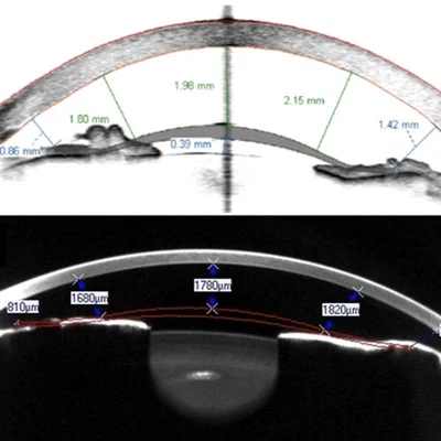
Our MRI protocols enable accurate three-dimensional reconstruction of individual eye geometry, forming the basis for patient-specific optical modelling. Using ray tracing simulations, we derive quantitative optical characteristics from these models: an approach that bridges ocular imaging and visual optics research.
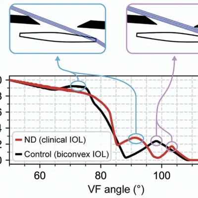
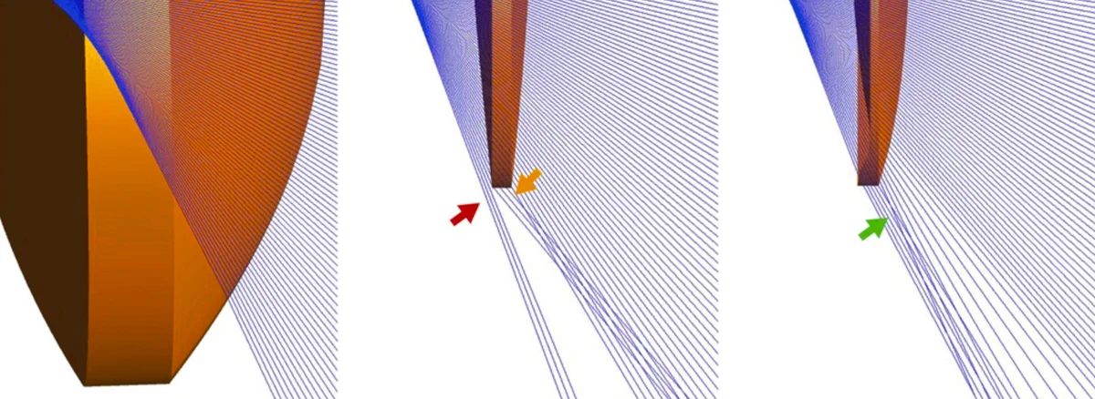
A key finding from this work is the anatomical explanation for Negative Dysphotopsia: our simulations demonstrated that peripheral visual loss after intraocular lens implantation results from light rays failing to reach the retina due to the geometry of the lens edge. Current work extends this framework to map fundus photograph coordinates onto 3D retinal anatomy, enabling more precise target delineation in ocular radiotherapy planning.
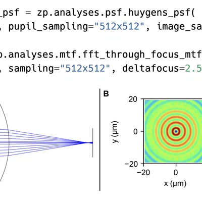
To support reproducible optical simulation in ophthalmology research, we developed Visisipy, an open-source Python package for ocular ray tracing, built for integration into standard scientific workflows.