Ocular Proton Therapy: Clipless MRI-Guided Treatment Planning
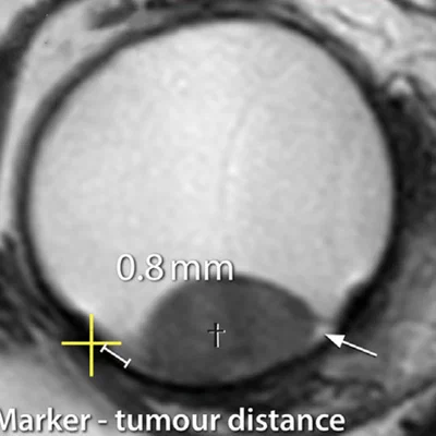
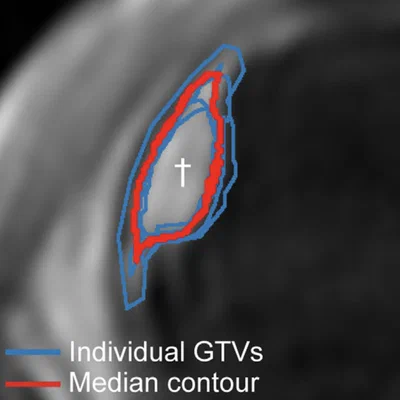
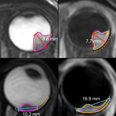
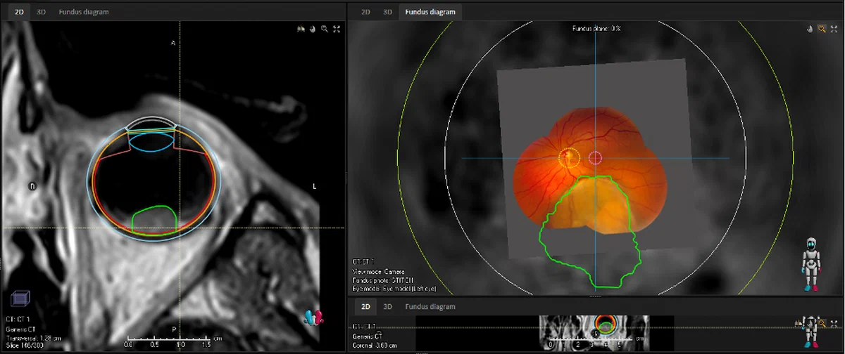
Conventional ocular proton therapy planning is based on a mathematical eye and tumour model derived from 2D ultrasound, with tantalum clips sutured to the eye wall as reference markers. We are replacing this approach with MRI-guided treatment planning, using three-dimensional high-resolution MR images to enable more conformal dose delivery — directly targeting the tumour while sparing critical visual structures. Meeting the established 98% local control benchmark remains a central requirement driving the precision of our methods.
In parallel, we are developing clipless ocular proton therapy in collaboration with HollandPTC, eliminating the surgical clip procedure and reducing both patient burden and treatment costs. Through patient preference studies conducted with Stichting Melanoom, we are further working to ensure that the added flexibility of MRI-based planning translates into genuinely patient-centred care.