Quantitative assessment of perfusion-weighted magnetic resonance imaging for the differential diagnosis of various intraocular lesions
Mulder, Vu, Ferreira, Klaassen and Beenakker
Research Topics
Abstract
Purpose: To conduct an exploratory analysis of quantitative and semi-quantitative perfusion-weighted MRI (PWI) parameters across various intraocular lesions and assess their relationship with fluorescein angiography (FAG) features.
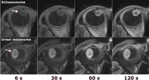
Methods: Thirty-eight patients with twelve different diagnoses who underwent 3T dynamic contrast-enhanced MRI (DCE-MRI) of the eye were included, with diagnosis confirmed either by histopathology or clinical course of the disease. Tofts pharmacokinetic modelling was used to calculate Ktrans, ve, and kep values. Semi-quantitative relative peak intensity and outflow percentage were derived from time-intensity curves and compared with quantitative parameters. FAG images were evaluated for blocking effects, pinpoint and diffuse leakages. Median values of quantitative and semi-quantitative parameters varied across lesion types.
Purpose: Haemangiomas showed the highest Ktrans (median = 6.35 min− 1) and kep (median = 3.8 min− 1). Uveal melanomas showed high Ktrans (median = 0.68 min− 1) and kep (median = 2.45 min− 1) in comparison with choroidal naevi and metastases. Quantitative and semi-quantitative parameters showed significant correlation across all intensity groups (all R2 > 0.36, all p < 0.04). The correlation between Ktrans and relative peak intensity differed for hypo- and isointense lesions versus hyperintense lesions (both p < 0.002). Statistical comparison between individual diagnoses was not possible due to limited sample size per diagnosis. Quantitative PWI parameters showed correspondence with FAG features. This study provided quantitative and semi-quantitative PWI parameters for various intraocular lesions.
Conclusion: Semi-quantitative PWI parameters may provide comparable information on lesion vascularity when stratified by T1 signal intensity.
Open Resources
Open Data: The perfusion metrics, T1 values and FAG classification of individual patients are made available as supplementary materials.