Reproducibility of perfusion-weighted and diffusion-weighted MR imaging in patients with ocular tumours
Janssen, Ferreira, Vu, Beenakker and Klaassen
Research Topics
Abstract
Introduction: Diffusion-weighted (DWI) and perfusion-weighted MRI (PWI) can assess biological tumour characteristics of uveal melanoma (UM), the most common primary intraocular malignant tumour in adults. Recent studies propose that these techniques can aid in the differential diagnosis, provide prognostic information, and enable early assessment of treatment response following radiation therapy. However, it is important to determine the extent to which any observed differences can be attributable to true biological changes in the tumour rather than day-to-day physiological or measurement variations. Therefore, this study aims to assess the reproducibility of ocular DWI and PWI measurements between two centres.
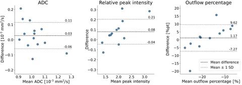
Methods: In this prospective study, 15 patients with UM were included. Each patient underwent two MRIs at two different centres with a mean interval of 28 days (range: 12-43 days). Apparent diffusion coefficient (ADC), relative peak intensity and outflow percentage of the tumour were compared between the two timepoints. Mean absolute differences between the two timepoints were calculated.
Results: Mean absolute differences between the two timepoints were 0.07 × 10⁻³ mm²/s (95% CI, 0.04-0.11) for ADC, 0.11 (95% CI, 0.04-0.18) for relative peak intensity and 6 (95% CI, 3-10) percentage points for outflow percentage.
Conclusion: This study shows that DWI and PWI parameters demonstrate reproducibility in UM patients between two different centres, with variabilities smaller than differences typically observed in differential diagnosis or treatment response assessments.
Open Resources
Open Data: The perfusion and diffusion metrics as well as MRI-based prominence measurements of both centers of all patients are made available as supplementary materials.
This manuscript is part of the PhD thesis Image-based ocular proton therapy by Lisa Klaassen.