Correction Method for Optical Scaling of Fundoscopy Images: Development, Validation, and First Implementation
Pors, Haasjes, Vught, Hoes, Luyten, Rijn, Vu, Rasch, Horeweg and Beenakker
Research Topics
Abstract
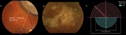
Purpose: Although fundus photography is extensively used in ophthalmology, refraction prevents accurate distance measurement on fundus images, as the resulting scaling differs between subjects due to varying ocular anatomy. We propose a PARaxial Optical fundus Scaling (PAROS) method to correct for this variation using commonly available clinical data.
Methods: The complete optics of the eye and fundus camera were modeled using ray transfer matrix formalism to obtain fundus image magnification. The subject’s ocular geometry was personalized using biometry, spherical equivalent of refraction (RSE), keratometry, and/or corneal topography data. The PAROS method was validated using 41 different eye phantoms and subsequently evaluated in 44 healthy phakic subjects (of whom 11 had phakic intraocular lenses [pIOLs]), 29 pseudophakic subjects, and 21 patients with uveal melanoma.
Results: Validation of the PAROS method showed small differences between model and actual image magnification (maximum 3.3%). Relative to the average eye, large differences in fundus magnification were observed, ranging from 0.79 to 1.48. Magnification was strongly inversely related to RSE (R2 = 0.67). In phakic subjects, magnification was directly proportional to axial length (R2 = 0.34). The inverse relation was seen in pIOL (R2 = 0.79) and pseudophakic (R2 = 0.12) subjects. RSE was a strong contributor to magnification differences (1%-83%). As this effect is not considered in the commonly used Bennett-Littmann method, statistically significant differences up to 40% (mean absolute 9%) were observed compared to the PAROS method (P < 0.001).
Conclusions: The significant differences in fundus image scaling observed among subjects can be accurately accounted for with the PAROS method, enabling more accurate quantitative assessment of fundus photography.
Open Resources
Open Materials: The PAROS methodology for calibrating fundus photographs is made available on GitHub.