A Comparison of 3 T and 7 T MRI for the Clinical Evaluation of Uveal Melanoma
Tang, Jaarsma‐Coes, Ferreira, Fonk, Marinkovic, Luyten and Beenakker
Research Topics
Abstract
Purpose: Magnetic resonance imaging (MRI) is increasingly being used in the diagnosis and treatment planning of uveal melanoma (UM), the most common primary intraocular tumor. Initially, 7 T MRI was primarily used, but more recently these techniques have been translated to 3 T, as it is more commonly available. In this study we compared the diagnostic performance of 3 T and 7 T MRI of UM.
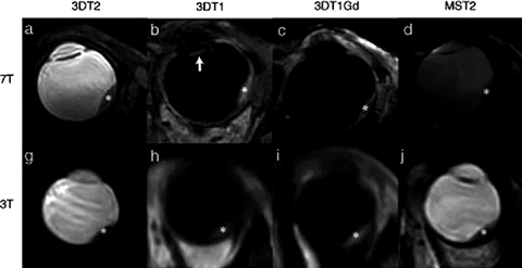
Methods: Twenty‐seven UM patients (19% female). 3 T: T1‐ and T2‐weighted three‐dimensional (3D) spin echo (SE) and multi‐slice (MS) SE, 7 T: T1‐weighted 3D gradient echo (GE), T2‐weighted 3D SE and MS SE, 3 T and 7 T GE dynamic contrast‐enhanced. T1 weighted images: acquired before and after Gadolinium (Gd) administration. For all sequences, scan and diagnostic quality was quantified using a 5‐point Likert scale. Signal intensities on T1 and T2 relative to choroid and eye muscle respectively were assessed as well as the tumor prominence. Finally, the perfusion time‐intensity curves (TICs) were classified as plateau, progressive, or wash‐out. Image quality scores were compared between both field strengths using Wilcoxon signed‐rank and McNemar tests. Paired t‐tests and Bland–Altman were used for comparing tumor prominences.
Results: Image quality was comparable between 3 T and 7 T, for 3DT1, 3DT2, 3DT1Gd (P = 0.86; P = 0.34; P = 0.78, respectively) and measuring tumor dimensions (P = 0.40). 2DT1 and 2DT2 image quality were rated better on 3 T compared to 7 T. Most UM had the same relative signal intensities at 3 T and 7 T on T1 (17/21) and T2 (13/17), and 16/18 diagnostic TICs received the same classification. Tumor prominence measurements were similar between field strengths (95% confidence interval: −0.37 mm to 0.03 mm, P = 0.097).
Conclusion: Diagnostic performance of the evaluated 3 T protocol proved to be as capable as 7 T, with the addition of 3 T being superior in assessing tumor growth into nearby anatomical structures compared to 7 T.