Eye-specific quantitative dynamic contrast-enhanced MRI analysis for patients with intraocular masses
Jaarsma-Coes, Ferreira, Houdt, Heide, Luyten and Beenakker
Research Topics
Abstract
Purpose: Dynamic contrast enhanced (DCE)-MRI is currently not generally used for intraocular masses as lesions are small, have an inhomogeneous T1 and the eye is prone to motion. The aim of this paper is to address these eye-specific challenges, enabling accurate ocular DCE-MRI.
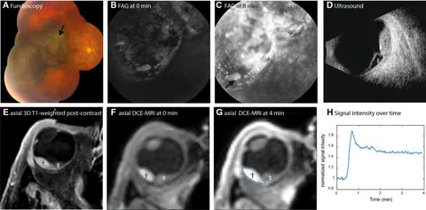
Methods: DCE-MRI of 19 uveal melanoma (UM) patients was acquired using a fat-suppressed 3D spoiled gradient echo sequence with TWIST (time-resolved angiography with stochastic trajectories sequence). The analysis consisted of a two-step registration method to correct for both head and eye motion. A T1 map was calculated to convert signal intensities to concentrations. Subsequently, the Tofts model was fitted voxel wise to obtain Ktrans and ve.
Results: Registration significantly improved the concentration curve quality (p < 0.001). The T1 of melanotic lesions was significantly lower than amelanotic lesions (888 ms vs 1350 ms, p = 0.03). The average achieved B1+ in the lesions was 91%. The average Ktrans was 0.46 min−1 (range 0.13–1.0) and the average ve was 0.22 (range 0.10–0.51).
Conclusion: Using this eye-specific analysis, DCE of intraocular masses is possible which might aid in the diagnosis, prognosis and follow-up of UM.
Open Resources
Open Data: The perfusion metrics and T1 values of individual patients are made available as supplementary materials.
This manuscript is part of the PhD thesis MRI for planning and characterization of uveal melanoma patients treated with proton beam therapy by Myriam Jaarsma-Coes.