Magnetic resonance imaging reveals possible cause of diplopia after Baerveldt glaucoma implantation
Islamaj, Vught, Jordaan-Kuip, Vermeer, Ferreira, Waard, Lemij and Beenakker
Research Topics
Abstract
Purpose: To assess if ocular motility impairment, and the ensuing diplopia, after Baerveldt Glaucoma device (BGI) implantation, is related to the presence of a large fluid reservoir (bleb), using Magnetic Resonance Imaging (MRI).
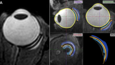
Methods: In a masked observational study (CCMO-registry number: NL65633.058.18), the eyes of 30 glaucoma patients with (n = 12) or without diplopia (n = 18) who had previously undergone BGI implantation were scanned with a 7 Tesla MRI-scanner. The substructures of the BGI-complex, including both blebs and plate, were segmented in 3D. Primary outcomes were a comparison of volume and height of the BGI-complex between patients with and without diplopia. Comparisons were performed by using an unpaired t-test, Fisher’s Exact or Mann-Whitney test. Correlations were determined by using Spearman correlation.
Results: The median volume and height of the BGI-complex was significantly higher in patients with compared to patients without diplopia (p = 0.007 and p = 0.025, respectively). Six patients had an excessively large total bleb volume (median of 1736.5mm3, interquartile range 1486.3–1933.9mm3), four of whom experienced diplopia (33% of the diplopia patients). Fibrotic strands through the BGI plate, intended to limit the height of the bleb, could be visualized but were not related to diplopia (75% versus 88%; p = 0.28).
Conclusion: With MRI, we show that in a significant number of diplopia cases a large bleb is present in the orbit. Given the large volume of these blebs, they are a likely explanation of the development of diplopia in at least some of the patients with diplopia after BGI implantation. Additionally, the MR-images confirm the presence of fibrotic strands. As these strands are also visible in patients with a large bleb, they are apparently not sufficient to restrict the bleb height.
Open Resources
Open Data: The MRI metrics (eg. bleb volume) and ophthalmic data (eg. ocular motility restrictions) of all patient can be found in the supplementary materials.