Measuring eye deformation between planning and proton beam therapy position using magnetic resonance imaging
Jaarsma-Coes, Marinkovic, Astreinidou, Schuurmans, Peters, Luyten, Rasch and Beenakker
Research Topics
Abstract
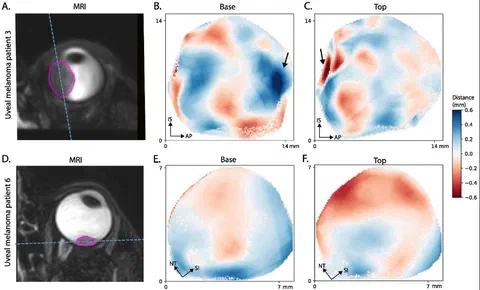
Purpose: Proton beam therapy (PBT) for uveal melanoma (UM) is performed in sitting position, while the acquisition of the Magnetic resonance (MR)-images for treatment planning is performed in supine position. We assessed the effect of this difference in position on the eye- and tumour- shape.
Methods: Seven subjects and six UM-patients were scanned in supine and a seating mimicking position. The distances between the tumour/sclera in both positions were calculated.
Results: The median distance between both positions was 0.1 mm.
Conclusion: Change in gravity direction produced no substantial changes in sclera and tumour shape, indicating that supinely acquired MR-images can be used to plan ocular-PBT.
This manuscript is part of the PhD thesis MRI for planning and characterization of uveal melanoma patients treated with proton beam therapy by Myriam Jaarsma-Coes.