Automated Retinal Topographic Maps Measured With Magnetic Resonance Imaging
Beenakker, Shamonin, Webb, Luyten and Stoel
Research Topics
Abstract
Purpose: Recent studies on ocular shape have raised increased interest in the peripheral characteristics of the eye, as it potentially triggers changes in the central vision. Current techniques are, however, not capable of accurately measuring the three-dimensional shape of the retina. We describe a new magnetic resonance imaging (MRI)-based method to obtain the retinal shape with high precision and use it to assess if differences in retinal shape could explain previously described trends in peripheral refraction.
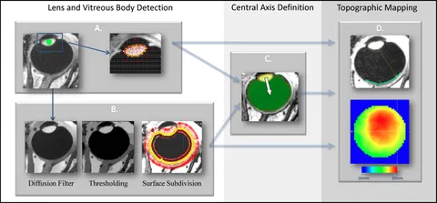
Methods: Twenty-one healthy subjects were examined using high-field ocular MRI. The resulting data were automatically segmented and processed to calculate the retinal topographic map. We validated the method against partial coherence interferometry and assessed the reproducibility for four subjects.
Results: The retinal topographic maps describe the retinal shape with subpixel reproducibility (SD between sessions = 0.11 mm). Comparison with partial coherence interferometry showed a mean difference of 0.08 mm, 95% confidence interval -0.39 to 0.55 mm, with a standard deviation of 0.23 mm. The data give a possible geometric explanation for the previously described trend in myopic eyes toward relatively hyperopic refraction in the periphery, with full three-dimensional information. The retinal maps furthermore show small, submillimeter, irregularities that could have an important influence on the subjects’ peripheral vision.
Conclusions: The possibility to quantitatively characterize the full three-dimensional retinal shape by MRI offers new ophthalmologic possibilities, such as quantitative geometric description of staphyloma. It could in addition be used as a validation technique, independent of standard optical methods, to measure the peripheral retinal shape.