High‐resolution MRI of uveal melanoma using a microcoil phased array at 7 T
Beenakker, Rijn, Luyten and Webb
Research Topics
Abstract
Purpose: High‐field MRI is a promising technique for the characterisation of ocular tumours, both in vivo and after enucleation.
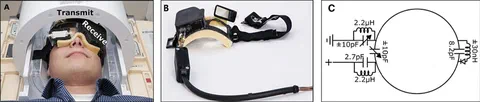
Methods: For in vivo imaging at 7 T, a dedicated three‐element microcoil array was constructed as a high‐sensitivity receive‐only device.
Results: Using a dedicated blink/fixation protocol, high‐resolution in vivo images could be acquired within 3 min in volunteers and patients with no requirement for post‐acquisition image registration. Quantitative measures of axial length, aqueous depth and lens thickness in a healthy volunteer were found to agree well with standard ocular biometric techniques. In a patient with uveal melanoma, in vivo MRI gave excellent tumour/aqueous body contrast. Ex vivo imaging of the enucleated eye showed significant heterogeneity within the tumour.