Christmas greeting

As we reflect on the past year, we celebrate the exciting scientific achievements that have shaped our work—some of which are subtly referenced in the picture above. Even more, we cherish the diverse and inspiring interactions we’ve shared with clinicians, (basic) scientists, patients, and industry partners.
Thank you for your invaluable contributions to our journey!
Click to discover the hidden references!
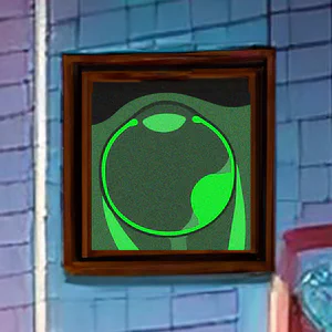
Uveal Melanoma
Most of our science is related to Uveal Melanoma, the most common form of eye cancer. This year we demonstrated that perfusion weighted MRI serves as an early biomarker of treatment response, providing crucial reassurance to patients.
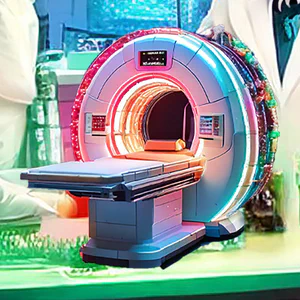
Magnetic Resonance Imaging
This year, we extended our MRI methods to other types of intra-ocular masses, making it a powerful tool in diagnosis when ophthalmic evaluations are inconclusive. In addition, we assisted multiple centers with the implementation of our MRI protocols.

Ocular Proton Beam Therapy
Its our ambition to preserve more of the patient’s vision after ocular radiotherapy. This year we have made important steps to further incorporate MRI in ocular proton therapy planning, enabling a more conformal treatment.
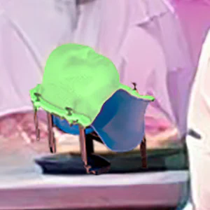
Clipless Ocular PBT
One of our larger projects is the development of clipless ocular PBT. We have finished the safety assessments and documentation of our clipless positioning system and are eagerly waiting for the start of the clinical trial in Q1 2025!
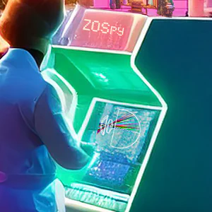
Ray tracing simulations
We extensively use ZOSPy for optical simulations to study vision and to combine fundus and MR-images. Next month, we will release ZOSPy 2.0, with many new features, whose development was funded through an NWO Open Science grant.
Evaluation of clinical results
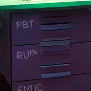
While developing new technologies is essential, evaluating and reporting on current practices is equally important. We therefore conducted an evaluation of the 820 UM patients treated with ruthenium-106 brachytherapy between 2012 and 2019. This study received ESTRO’s “Best Junior Presentation” Award.

Ocular Cone-Beam CT Scanner
On January 1st, we will start our project to develop an ocular cone-beam CT scanner as an accessible 3D imaging modality for the eye and orbit, with direct applications for ocular proton therapy.

Proton spin
Protons are central to most of our projects. They not only generate the MRI signal, they are also particles we use to irradiate tumours.
We wish you a positive 2025!